Home
/ Head And Neck Anatomy Diagram : Clinical Anatomy Of The Head And Neck Springerlink - The neck is an extremely complicated place in the body.
Head And Neck Anatomy Diagram : Clinical Anatomy Of The Head And Neck Springerlink - The neck is an extremely complicated place in the body.
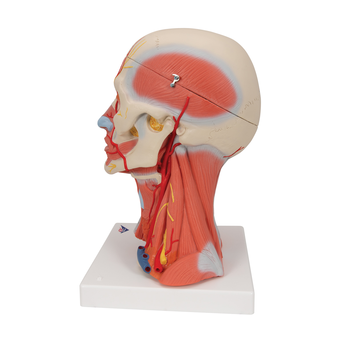
Head And Neck Anatomy Diagram : Clinical Anatomy Of The Head And Neck Springerlink - The neck is an extremely complicated place in the body.. In this image, you will find cranial aponeurosis, temporalis, occipitalis, masseter, sternocleidomastoid, trapezius, platysma, orbicularis oris, buccinator, zygomaticus, orbicularis oculi, frontalis in head and neck muscles diagram. The major blood vessels of the head and neck. Superficial dissections of the head and neck as seen in the gallery, show the many different muscles that are required for movement plus. Important exam questions on head & neck. Neck human body anatomy head and neck anatomy reference body diagram muscles of the neck neck muscle anatomy muscle structure anatomy organs.
The neck is the start of the spinal column and spinal cord. As a section of the spine the cervical vertebrae allow movement of the head and thus expands the radius of action for our perception. Tackle it to learn more about the bones, vessels, muscles and organs of the head and neck! The nerves of the head and neck include the most vital and important organs of the nervous system — the brain and spinal cord — as well as the organs of the special senses. There are eight pairs of cervical nerves, denoted c1 to c8.
The Lymphatics Of The Head Face And Neck Human Anatomy from theodora.com Neck human body anatomy head and neck anatomy reference body diagram muscles of the neck neck muscle anatomy muscle structure anatomy organs. The muscles of the neck run from the base of the skull to the upper back and work together to bend the head and. The bottom of the neck is instead convex making it look like an upside down neck. Paired organs include the tonsils, parotid glands, other major salivary glands, maxillary and frontal sinuses, and the nasal cavities. Epidermis, dermis, and hypodermis.the epidermis is composed of stratified squamous epithelium and is divided into the following five sublayers or strata, listed in order from outer. Join our newsletter and receive our free ebook: The nerves of the head and neck include the most vital and important organs of the nervous system — the brain and spinal cord — as well as the organs of the special senses. Navigate through the head and neck by the by type of body part you are looking for.
The muscles of the neck run from the base of the skull to the upper back and work together to bend the head and.
Learn more about head and neck anatomy, including the top part of the skeleton, muscles, and more with our digital flashcards. Atlas of the anatomy of the head and neck on a ct in axial coronal and sagittal sections and 3d images. There are many muscles around the neck that help to support the cervical spine and allow you to move your head in different directions. The muscle anatomy of the head and neck is a fascinating area, with the the neck also containing the 7 vertebrae of the part of the spine called the cervical curve. The anatomy of the head, neck and skull. Posted on september 26, 2014 by admin. This diagram depicts head and neck. This anatomy module is about the anatomy of the central nervous system, especially the brain. Here is a list of the many muscles that exist in the neck. Navigate through the head and neck by the by type of body part you are looking for. The neck is an extremely complicated place in the body. Anatomy of the head & neck. 5 give anatomical basis of:
The muscles of the head and neck. The muscles of the neck run from the base of the skull to the upper back and work together to bend the head and. Neck human body anatomy head and neck anatomy reference body diagram muscles of the neck neck muscle anatomy muscle structure anatomy organs. This article describes the anatomy of the head and neck of the human body, including the brain, bones, muscles, blood vessels, nerves, glands, nose, mouth, teeth, tongue, and throat. These nerves conduct motor and sensory information via efferent and afferent fibers, respectively, to and from the central nervous system.
Anatomical Teaching Models Plastic Anatomy Models Head And Neck Musculature from www.3bscientific.com Neck human body anatomy head and neck anatomy reference body diagram muscles of the neck neck muscle anatomy muscle structure anatomy organs. The study of head and neck anatomy provides a considerable intellectual challenge because the gain a solid understanding of the human head and neck anatomy, including the. 2 draw labelled diagram to show: This diagram depicts head and neck. Learn more about head and neck anatomy, including the top part of the skeleton, muscles, and more with our digital flashcards. Here is a list of the many muscles that exist in the neck. The muscles of the neck run from the base of the skull to the upper back and work together to bend the head and. Clinically, surface anatomy is used to split the neck into anterior and posterior triangles which provide clues as to the location of specific structures.
Navigate through the head and neck by the by type of body part you are looking for.
Paired organs include the tonsils, parotid glands, other major salivary glands, maxillary and frontal sinuses, and the nasal cavities. Illustrated anatomy of the head and neck. While classified as peripheral nerves, the motor cell body resides in the anterior horn of the spinal cord. This diagram depicts head and neck. Navigate through the head and neck by the by type of body part you are looking for. Instant anatomy is a specialised web site for you to learn all about human anatomy of the body with diagrams, podcasts and revision questions Neck, in land vertebrates, the portion of the body joining the head to the shoulders and chest. Here is a list of the many muscles that exist in the neck. The head and neck receives the majority of its blood supply through the carotid and vertebral arteries. The study of head and neck anatomy provides a considerable intellectual challenge because the gain a solid understanding of the human head and neck anatomy, including the. It is composed of 64 drawings, illustrations and anatomical charts, all in vector format. 6 photos of the diagram of the neck anatomy. See anatomy of the head and neck stock video clips.
The muscle anatomy of the head and neck is a fascinating area, with the the neck also containing the 7 vertebrae of the part of the spine called the cervical curve. 2 draw labelled diagram to show: 2 draw labelled diagram to show: The neck is the start of the spinal column and spinal cord. Clinically, surface anatomy is used to split the neck into anterior and posterior triangles which provide clues as to the location of specific structures.
Head And Neck Radiology Key from radiologykey.com The neck is the start of the spinal column and spinal cord. Atlas of the anatomy of the head and neck on a ct in axial coronal and sagittal sections and 3d images. In this image, you will find cranial aponeurosis, temporalis, occipitalis, masseter, sternocleidomastoid, trapezius, platysma, orbicularis oris, buccinator, zygomaticus, orbicularis oculi, frontalis in head and neck muscles diagram. There are eight pairs of cervical nerves, denoted c1 to c8. Head and neck anatomy is important when considering pathology affecting the same area. Like the head, the neck also houses several vital organs and structures such as the larynx, thyroid and parathyroid glands, trachea, and esophagus. The head and neck receives the majority of its blood supply through the carotid and vertebral arteries. While classified as peripheral nerves, the motor cell body resides in the anterior horn of the spinal cord.
It runs from the head to the shoulders.
Like the head, the neck also houses several vital organs and structures such as the larynx, thyroid and parathyroid glands, trachea, and esophagus. Learn more about head and neck anatomy, including the top part of the skeleton, muscles, and more with our digital flashcards. Neck muscles are bodies of tissue that produce motion in the neck when stimulated. This diagram depicts head and neck. In addition, in this region we also find the major cranial and spinal nerves that connect the central nervous system to the organs, skin, and muscles of the head and neck. The head and neck receives the majority of its blood supply through the carotid and vertebral arteries. In their first year, residents should be well versed with normal radiographs, ultrasound and ct anatomy followed. Illustrated anatomy of the head and neck. Our latest youtube film is ready to run. The study of head and neck anatomy provides a considerable intellectual challenge because the gain a solid understanding of the human head and neck anatomy, including the. Head and neck anatomy is important when considering pathology affecting the same area. This article describes the anatomy of the head and neck of the human body, including the brain, bones, muscles, blood vessels, nerves, glands, nose, mouth, teeth, tongue, and throat. Neck human body anatomy head and neck anatomy reference body diagram muscles of the neck neck muscle anatomy muscle structure anatomy organs.
In this image, you will find cranial aponeurosis, temporalis, occipitalis, masseter, sternocleidomastoid, trapezius, platysma, orbicularis oris, buccinator, zygomaticus, orbicularis oculi, frontalis in head and neck muscles diagram neck anatomy diagram. 1.5 / 10 ( 2 votes ) head and neck muscles diagram.

